Microtomes are vital instruments in the pathology department because they enable the accurate cutting of tiny tissue slices for microscopic examination. A microtome can supply you with the high-quality samples you need for your research, whether you’re examining the structure of cells or a tissue’s characteristics or diagnosis of a disease. Another essential device in the pathology lab is the tissue processor.
Let’s delve into the world of microtomes and learn about the numerous kinds of microtomes that are available, their history, how they function, and the various uses that they have in the scientific and medical fields. This article will provide you with a thorough introduction to this intriguing equipment, whether you’re a researcher, a student, or just interested in the technology underlying microtomes. Now let’s get going and discover more about the astounding accuracy of microtomes.
What is a microtome?
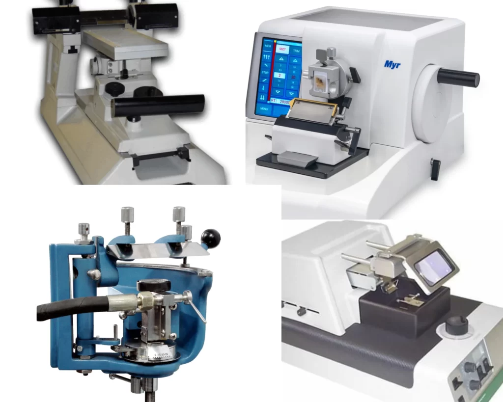
A microtome is a unique scientific tool used to slice or section thin biological material for microscopic analysis. It enables scientists to look within tissues and cells to spot any structural reforms or disorders. In histology, pathology, and neuroscience research, microtomes are frequently employed to create tissue sections for dyeing and microscopic inspection. Microtomes come in a variety of forms, such as rotary, cryo, vibratome, sledge, freezing, and laser microtomes. The kind of tissue being sectioned, the thickness of sections needed, and the caliber of the sections produced are the criteria for selecting a microtome for the sectioning of tissues.
History of Microtomes- How it all began
The development of microtomes dates back to the 17th century. It was believed that studying thin sections of tissues could offer significant knowledge about the makeup and function of living organisms at the time, thus scientists and researchers began looking for techniques to create thin slices of tissues for microscopic investigation.
George Adams, a British optician, developed the first microtome in 1770. It was a straightforward tool that made it possible to slice thin pieces of ivory or other hard materials for microscopic examination. But it wasn’t until the early 1800s that a Frenchman, Louis Odier, created the very first microtome that could section soft tissues. Take a look at how the video otoscope work.
The first tool capable of slicing soft tissues for microscopic observation was Louis Odier’s microtome, developed during the early 1800s. It allowed for accurate and equal sectioning. It relied on a wedge-shaped knife propelled by a threaded rod. Unfortunately, it had shortcomings, such as the inability to cut extremely thin portions.
Wilhelm was the next scientist to make an improvement in the development of microtomes. Wilhelm His Sr., a German-based Swiss anatomist, developed the very first microtome in 1865. His microtome was a razor blade on a gliding carriage that could be moved over a stationary object to cut thin slices with accuracy and consistency. This early microtome required a lot of manual work and was time-consuming to operate.
The use of technology has led to the creation of all sorts of microtomes including automated ones. Microtomes are now utilized in a variety of scientific fields, including histology, pathology, and neuroscience. They come in a variety of shapes and sizes. Current microtomes may create slices as flat as 2 microns and employ a variety of cutting edges, including diamond knives, glass knives, and disposable blades. this is how RA660 audiometer is revolutionizing hearing test.
Types of Microtomes and what they are used for
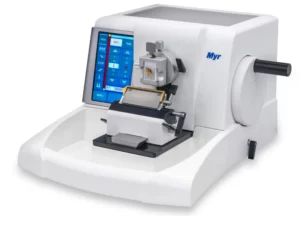
- Rotary microtomes- The most popular kind of microtome is the rotary microtome. To cut thin slices, a circular blade is revolved around a stationary object. The sections produced by rotary microtomes are uniformly thin and straightforward to utilize. They are perfect for slicing through animal tissues, plant sections, and tissues encased in paraffin.
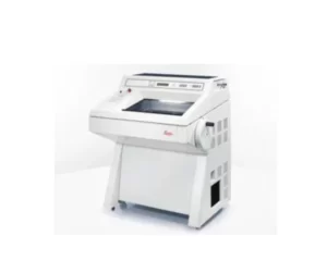
- Cryo-microtomes-They slice frozen specimens into thin parts using a chilled cutting platform and a sharp blade. In pathology labs, cryo-microtomes are frequently used to analyze fresh tissues and generate quality sections of unfixed, fresh specimens.
- Vibratome microtomes– They are used by vibratome microtomes to slice soft tissues into tiny pieces. They are frequently used in neuroscience research to prepare brain sections for electrophysiological tests because they are perfect for cutting slices of fresh, unfixed tissues.

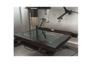
- Laser microtomes- They use a laser beam to slice away tiny pieces of either soft or hard tissues. They enable the accurate cutting of small sections and are frequently utilized in the imaging of biological tissues.
- Freezing microtomes- A cold stage is used in freezing microtomes to freeze the specimen, after which tiny slices are cut with a razor blade. They can provide excellent sections of unfixed tissues and are frequently employed in plant biology to investigate fresh tissues.

- Sledge microtomes- They large, powerful microtomes can be used to slice through substantial pieces of tough materials like bone, and teeth.

Conclusion
The development of microtomes is proof of the value of innovation in scientific study. The instrument has experienced several design and functional developments, allowing for increased precision and accuracy in sectioning. From the initial periods of employing hand-held scalpels to the invention of automated microtomes. Microtomes are now fundamental pieces of equipment in histology labs all over the world. They are used for a wide range of tasks, such as the analysis of plant and animal tissues. Microtomes are anticipated to continue to be crucial tools in scientific investigations for many years to come as technology gets better.