In histology and pathology research, thin tissue sections are often cut with the help of a sliding microtome, a precision cutting instrument. It works by moving a tissue sample block against a cutting edge, producing thin pieces of tissue that are uniform and consistent. A thorough examination of tissue architecture and cellular constituents is possible due to the microtome’s ability to create chunks as tiny as a few microns in size. Staining, immunohistochemistry, and electron microscopy are only a few of the uses for the generated sections. Researchers can get insight into the intricate structures and processes of living creatures using the sliding microtome, which is a crucial instrument in many fields. Read about the electric dental handpiece
What is a sliding microtome?
A sliding microtome is a scientific tool used to section tissue samples into thin chunks for microscopic analysis in histology, neurology and pathology. A tissue sample is cut into small tissue sections using the sliding microtome to operate. The platform on which the block is attached slides back and forth along a guide rail. A sled fitted with a razor blade—typically composed of steel or diamond—moves horizontally across the platform, slicing the tissue as it does so. The location and angle of the blade and the distance the tissue holder travels with each slice can be changed to alter the thickness of the sections. Take a look at the cryostat microtome.
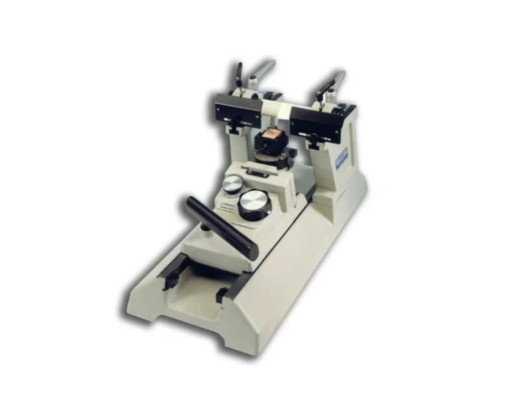
Parts of a sliding microtome
- Handle– Controls the movement of the tissue holder for the slicing of the tissue.
- Tissue holder- The tissue is fixed in the tissue holder for sectioning.
- Blade holder– It holds the blade firmly in position to slice the tissues.
- Feed mechanism- It ensues the advancement od the tissue for sectioning.
- Coarse adjustment angle- It alters the angle of the blade for precise sectioning.
- Micrometer adjustment- Controls the thickness of the tissue to be sectioned.
- Waste tray- Gathers debris of the tissues during sectioning.
- Safety features- It comes with guards and covers as safety precautions to shield the user from unintentional harm.
What is the principle of the Sliding Microtome?
The sliding microtome utilizes the idea of slicing thin pieces of tissue samples for microscopic analysis with the aid of a sharp blade. The blade of the sliding microtome is static whereas the tissue sample is moved through it in tiny steps. The knife is held in a blade holder that is set at a specific angle, and the specimen tissue sample is kept stationary on a tissue holder. The blade holder is usually adjusted to enable the blade’s angle to be changed to improve sectioning of the tissues. Read about the principle of the rotary microtome.
A tiny piece of the tissue is cut off as it is pushed over the blade. By altering the space between the blade and the tissue as well as the speed that the tissue is carried over the blade, the thickness of the section can be altered. The resulting sections, which can be just a few micrometers thick, are ideal for microscopic study. Do not forget about dental composite.
How to operate the Sliding Microtome
- Set the thickness- Change the settings of the microtome to obtain your desired thickness.
- Insert the tissue- Fix the tissue into the tissue holder and ensure tat is properly positioned before you begin sectioning.
- Adjust the blade- Alter the position and angle of the blade to your preference for precise sectioning.
- Begin cutting- Push the handle of microtome to commence the sectioning of the tissues.
- Gather tissues- Collect tissues unto slides for staining and viewing under the microscope. Prior to this, tissues can be placed in a water bath at 37oC to straighten the edges.
Maintenance
- Cleaning- Non-abrasive sponge can be used to get rid of debris of tissues and grease. Detergent can be added to remove tough stains sticked to the microtome.
- Lubricating– Adding few drops of oil will prevent rusting and maintain the smooth operation of the moving parts of the microtome.
- Blade maintenance- Sharpen or replace blades when the cutting edge becomes blunt. This to prevent serration of tissues being sectioned.
- Calibration– Verify the accuracy and precision of the blades regularly by checking the consistency of the tissues. Alter the angle or position to get the appropriate size.
- Storage- keep the sliding microtome in a clean and dry area and cover it with a hood to prevent dust from gathering on it.
Conclusion
In biological study and medical diagnosis, sliding microtomes are essential tools. For the purpose of dissecting cellular and tissue architecture, they enable the accurate sectioning of tissues into thin, homogenous slices. The advantages of the sliding microtome exceed its disadvantages by a wide margin, notwithstanding the requirement for competence in using the device and the time-consuming nature of the slicing procedure. The sliding microtome has become an essential tool in many fields of scientific and medical research because of its capacity to produce high-quality tissue slices, and it is certain to keep playing a significant role in expanding our knowledge of the biological world. Do not forget to read about the microscope slide scanner.